FEMORAL ACETABULAR IMPINGEMENT (FAI)
Femoroacetabular impingement or FAI is a condition where the bones of your hip joint come too close and pinch tissue or cause too much friction. Usually, the ball of the hip joint (femoral head) sits on the femoral neck similar to an ice cream sitting on a cone. The pinching and friction occurs when the femoral head and neck contact the socket (acetabulum), creating damage to the hip joint. The pinching or friction may cause damage to the labrum (a fibrous cartilage that lines the outer edge of the socket) and/or the articular cartilage (the white covering over the bony surfaces that results in the very smooth surface gliding of the joint).
CAUSES
FAI generally occurs as two forms: Cam and Pincer. The Cam form describes the femoral head and neck relationship as aspherical or not perfectly round. This loss of roundness contributes to abnormal contact between the head and socket as the hip goes through a range of motion. The Pincer form describes the situation where there is overcoverage of the socket or acetabulum relative to the ball or femoral head. This over-coverage typically exists along the front-top rim of the socket (acetabulum). The end result is that the labral cartilage gets “pinched” between the rim of the socket and the front part where the femoral head meets the femoral neck. The Pincer form of the impingement is typically the results of “retroversion”, where the socket is pointed backwards a bit (rather than the usual situation where it is angled forwards), or where the socket is too deep. Very often, the Cam and Pincer forms exist together. The cause of these bony variations is not known at this time, but the cam lesion is likely related to activities performed during teenage years.
FAI is associated with articular cartilage damage and labral tears and may result in hip arthritis at a younger age than usually occurs.

Figure 1. Cam vs. Pincer type impingement.
WHO IS AT RISK
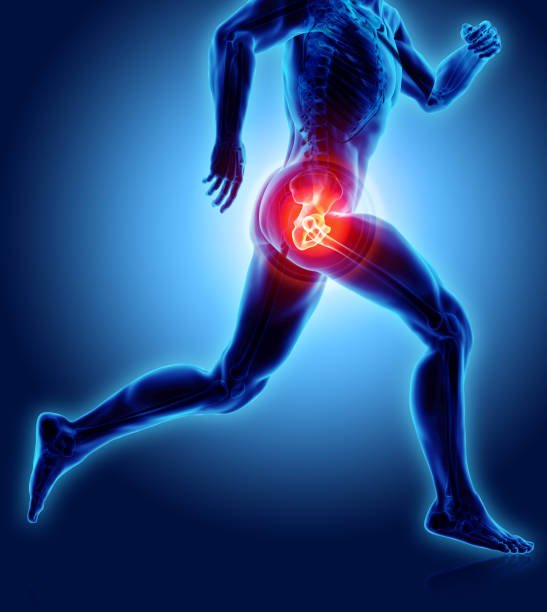
FAI is common in high level athletes, but also occurs in active individuals. I have patients with FAI who are involved in almost any / every sport. Sports particularly associated with FAI include Martial Arts, Ballet, Cycling, Rowing, Golf, Tennis, Soccer, Football, Ice Hockey, Baseball, Lacrosse, Field Hockey, Rugby, Water Polo, and Deep squatting activities such as power lifting.
SYMPTOMS OF FAI
There may be no pain or symptoms
Pain or aching (usually located at the inner hip, or groin area), usually after walking, or prolonged sitting (such as in a car)
A locking, clicking or “catching” sensation within the joint
Pain sitting for long periods of time, like in a car
Difficulty putting on your socks and/or shoes
Difficulty walking up hill
Low back pain
Pain at the SI (sacroiliac joint on back of pelvis), the buttock, or greater trochanter (side of hip)
It is often confused with other sources of pain, such as hipflexor tendinitis, pain from the back (disc or spine), testicular pain, sports hernia
EVALUATION
Your doctor will ask about your hip (your symptoms and how the pain started, for how long, etc) and perform an examination. Your doctor will move your hips and legs in different positions to assess your range of motion and evaluate the positions where your hip hurts.
To confirm a diagnosis you will likely get X-Rays of your hip. Plain X-Rays are very important in the evaluation of the active patient with hip pain. There is information the plain X-Rays provide that even MRI’s cannot, so even if you have an MRI, you will need an X-Ray. Often, you may undergo a special type of magnetic resonance imaging (MRI) called magnetic resonance arthrography (MRA).
Magnetic resonance arthrography (MRA) is a noninvasive, non-irradiating imaging technique that uses a magnetic field and radio waves to evaluate your hip. While X-Rays show bones well, the MRI is particularly good at showing the non-bony structures of the body, such as the labrum and articular cartilage. Further, while X-Rays are like looking at shadows, the MRI allows evaluation of the tissues around the hip in slices (like slices of bread as opposed to seeing the whole loaf without what is inside) and allows viewing from different views. During magnetic resonance arthrography, dye (contrast material) is injected into the joint space to help make images more clear. Frequently, local anesthetic (numbing medicine) is added to the contrast material to help determine if the pain is coming from inside the joint. The MRI will also help eliminate certain causes of non-FAI hip pain including avascular necrosis (dead bone) and tumors.
Sometimes your physician may order a CT or CAT scan. This study can help understand the exact shape of the bones of the hip, but is not essential to the diagnosis of FAI. 3-D CT scans are particularly good at giving the surgeon a very realistic perspective of the shape of the bone.
TREATMENT
The underlying problem with FAI is a bony abnormality. This bony shape will not change with physical therapy or rest. However, the shape of the bones itself do not cause pain. Other structures that can be injured with FAI, such as the labrum, or articular cartilage may cause the pain in the hip. Neither the labrum or articular cartilage have much capacity to heal, but sometimes these structures, even when injured do not cause pain or other symptoms. Thus, for those with symptoms the initial treatment may involve rest and rehabilitation, while those that have symptoms that persist, arthroscopic surgery may be needed. We have seen many high level athletes improve and return to high level sports with just physical therapy.
The long term sequelae of FAI has not been conclusively proven, but there is much evidence that it may be a major cause of premature arthritis of the hip. It has also not been proven that surgery for FAI will prevent arthritis. However, removing the offending bone may help reduce further injury to the joint, while also reducing symptoms. The results of surgery are clearly better when there is no articular cartilage damage. Thus, most physicians familiar with this problem often recommend early surgical intervention for symptomatic patients with FAI.
NON-OPERATIVE APPROACH
Nonoperative management of FAI is usually the first step in treatment. However, this route also involves a change in lifestyle from active to less active and a commitment to maintaining hip strength. A good physical therapy program focusing on hip and core strengthening instead of stretching may be beneficial. Stretching associated with yoga and sometimes physical therapy may make the symptoms worse. Activity modification should involve avoiding activities that take the hip through extreme or full ranges of motion. Anti-inflammatory medications can also be tried.
SURGICAL TREATMENT
Surgery for FAI can be performed using hip arthroscopy or open surgery. In hip arthroscopy, the hip is distracted and an arthroscope (a video-camera about the size of a pen) is used to look in the joint to see and treat damage that is found using two to five incisions that are about ¼” in size. Often, all of the components of FAI such as the labral tear, damaged articular cartilage, and bony changes between the ball and socket can be treated with the assistance of the arthroscope. Repair of a torn labrum as well as stimulating new cartilage growth (microfracture) are often possible with the arthroscopic approach. A hip arthroscopy is an outpatient procedure (go home the same day) and takes 1 – 2 hours.

Figure 2. Surgical Approach for FAI.
RECOVERY FROM SURGERY
The patient is on crutches after surgery. The length of time you are on crutches will depend on what is done and your surgeon’s preference. Recovery time from most FAI surgical procedures is 4 – 6 months to full, unrestricted activity. Your postoperative activity level will depend on your surgeon’s recommendation, the type of surgery performed, and the condition of the hip joint at the time of surgery.
Commonly Asked Questions About FAI
-
It seems Cam type FAI (the femoral head / ball being out of round) is related to sporting activities the patient performed while in their teenage years. No one knows how Pincer FAI (socket depth or rotation) occurs, whether it is from birth (congenital) or develops during periods of growth (acquired). However, even though you may have the bony changes of FAI, you may not have symptoms unless you participate in activities that require large hip motions or motions at the extremes
-
Some experts believe that significant athletic activity before skeletal maturity increases the risk of FAI, but no one really knows. Contact and collision sports (i.e., soccer, football) are associated with Cam impingement.
-
Not necessarily. However, first evaluation is done with x-rays which would show significant arthritis. MRI scans may show loss of articular cartilage, but frequently, there is significant loss of articular cartilage inside the hip that is not seen on MRI or X-ray.
-
At times, an MRI will be read as “Normal” but the clinical history, physical exam, and plain x-ray films indicate FAI. In this situation, further investigation with an arthroscopic surgery may be needed.
-
In general, most patients with hip pain due to labral tears or FAI often have seen multiple doctors and have significant delays in diagnosis before the correct diagnosis and treatment is instituted.
-
Hip labral tears are associated with FAI. If you have had your labral tear treated and are still having pain, you may have FAI. The success of labral surgery is much less if there is untreated FAI. Further, residual impingement may lead to further loss of hip cartilage.
-
Yes, it is possible for both hips to have FAI. However, while 85% of my patients have similar anatomy on both sides (X-rays of FAI on both sides), only 15 – 25% of my patients have symptoms on both sides needing surgery.
-
Some patients with FAI complain of stiffness and loss of hip range of motion without any significant pain. Progressive loss of motion in the hip can be associated with ongoing FAI. Speak to your physician about your options.
-
DDH or developmental dysplasia of the hip is a different diagnosis than FAI. DDH generally refers to too little coverage of the ball by the socket. FAI generally refers to too much coverage of the ball by the socket. Both DDH and FAI are associated with labral tears and articular cartilage damage. Some patients with DDH (shallow socket) can have cam FAI as well.
-
If one has a diagnosis of FAI or suspects FAI, one should be evaluated by an orthopaedic specialist who is adept in treating hip disorders. Your physician should have experience with either open surgical hip dislocation or hip arthroscopy.
-
Typically, FAI that produces symptoms for more than 2 months should be evaluated for surgical treatment. A longer wait may lead to further damage of the joint. We have found that those who wait longer between the onset of symptoms and surgery do not have as full a recovery / return to sports, as those that have symptoms of a shorter duration.
-
Generally, FAI is a chronic condition that does not typically respond well to hip injections or physical therapy over the long term. The injections may make the hip feel better for a short duration. However, a good physical therapy program focusing on hip strengthening instead of stretching may be beneficial. The key is that forced stretching may make the symptoms worse.
-
Non-operative treatment is always an option. If you follow a conservative treatment plan of active relative rest, stretching and strengthening, the pain and swelling may go down. If however, you have a labral tear or articular cartilage damage, these generally do not heal. Usually, the pain and swelling will return once you return to your chosen sporting activity.
-
The pain may come and go, but likely would not decrease significantly or for an extended period of time, especially if you continue with sporting activity, without surgical intervention.
-
Yes… The postoperative rehabilitation of a total hip replacement (total hip arthroplasty) is significantly shorter than an FAI procedure. However, hip replacements have a limited longevity, especially for younger patients. The same is true for a hip-resurfacing type of procedures. Both the resurfacing arthroplasty and the total hip arthroplasty involve removal of the damaged joint surfaces and replacement with man-made materials (i.e., metal, plastic, ceramic) which are subject to wear. The wear results in joint debris which may shorten the life of the replacement.
Once your hip replacement fails or wears out, your revision hip replacement does not last as long or work as well as your first and has a higher complication rate. And each subsequent replacement will not work as well or last as long as the one before and has a higher complication risk associated with it. As such, the goal is to put off having a joint replacement as long as possible, so that the one you finally get is the only one you need. Once you go down the path of hip replacement, there is no turning back.
-
Sedentary work can be resumed in one to two weeks. Labor intensive work maybe 12 – 20 weeks.
-
Once you have good control of your leg and you are not taking any narcotic medications. This is usually 1-2 weeks
-
Yes, though the amount and duration depends on what is done for your hip and your surgeon’s preferences. Usually Physical Therapy (PT) starts the week after surgery. Additionally, you will likely also need to perform home exercises after surgery.
-
Yes but you don’t need to. Spinal anesthesia is possible but general anesthesia is recommended to reduce complication risk.
-
The length of time you are put on crutches after FAI surgery depends on what procedures are done, and your surgeon’s experience. Your rehabilitation progress will determine the weaning process as well as the extent of the tear and/or associated problems.
-
Yes. The ability to detect articular cartilage injury before surgery still is not perfected, even with MRI. As hip arthroscopy techniques become more refined the incidence and ability to treat cartilage problems are both increasing. The presence of cartilage lesions (articular cartilage) is identified at the time of surgery and is treated by debridement (cleaning it up) and/or microfracture (where we poke holes in the bone to stimulate growth of a scar cartilage to replace the lost articular cartilage).
-
Surgery is done to treat your symptoms, usually groin pain, as well as to reduce worsening of the tear. There is no guarantee that a recurrent tear will not occur nor is there any guarantee that surgery will prevent arthritis. Recurrent tears are, however, unusual. Also, it is not known whether removing the torn cartilage will prevent further damage.
-
Complications from FAI hip surgery are uncommon but include the following:
DVT (blood clot) that may break off and go to the lungs (pulmonary embolus)
Infection
Bleeding
Femoral neck fracture (broken hip)
Avascular Necrosis (AVN) of the femoral head (dead bone)
Heterotopic ossification (abnormal bone formation in soft tissues)
Nerve injury
Sciatic or femoral nerve {leg numbness or weakness)
Lateral Femoral Cutaneous Nerve (LFCN) {numb outer thigh}
Pudendal {numbness in the groin / genitals}
Scarring/Adhesions
Continued Pain
Damage to the cartilage
Need for further surgery
Hip Instability
Not removing enough bone or too much bone -
All activities, even rolling over in bed can cause hip stresses. The most important exercises are ones which create normal flexibility about your hip and normal, protective strength. In some situations, activities that require extremes in your range of motion of your hip, may cause the pinching of tissues, resulting in damage to the torn labrum and articular cartilage.
-
Feeling worse after surgery is always a possibility, however, the likelihood of that is very small.
-
Pain medications can be ordered but are not recommended prior to surgery. Non-steroidal anti-inflammatory (Advil, Aleve, etc) and Tylenol mixed together are often better than either alone. However it is recommended that you not take anti-inflammatory medications for the 2 weeks prior to surgery.